Anatomic Terms allow us to describe the body clearly and precisely using planes, areas and lines. Instead of your doctor saying “his knee hurts” she can say “his knee hurts in the anterolateral region” and another doctor will know exactly what is meant. Below are some anatomic terms surgeons use as these terms apply to the hip:(1)
- Anterior — the abdominal side (front) of the hip
- Posterior — the back side of the hip
- Medial — the side of the hip closest to the spine
- Lateral — the side of the hip farthest from the spine
- Abduction — move away from the body (raising the leg)
- Adduction — move toward the body (lowering the leg)
- Proximal — located nearest to the point of attachment or reference, or center of the body
- Distal — located farthest from the point of attachment or reference, or center of the body
- Inferior — located beneath, under or below; under surface (2)

Bones
The hip is formed where the thigh bone (femur) meets the three bones that make up the pelvis: the ilium, the pubis (pubic bone) and the ischium. You can feel the arching bones of the ilium by placing your hands on your waist. The pubis attaches to the lower part of the ilium and curves forward. The ischium is slightly behind the pubis. The three bones converge to form the acetabulum, a deep socket on the outer edge of the pelvis.(1)
Joints
The hip joins the leg to the trunk of the body at the hip joint. The hip joint is made up of the ball of the femoral head that fits into the cup-shaped acetabulum. The large round head of the femur rotates and glides within the acetabulum. (1)
Ligaments
The stability of the hip is increased by the strong ligaments that encircle the hip. These ligaments completely encompass the hip joint and form the joint capsule.(1)
Tendons
Muscles
The muscles of the thigh and lower back work together to keep the hip stable, aligned and moving. It is the muscles of the hip that allow the 4 basic movements of the hip:
Muscles of the Hip Region:
Nerves (major)
Nerves carry signals from the brainto the muscles to move the hip and carries signals from the muscles back to the brain about pain, pressure and temperature.
Blood Vessels (Major)
Bursae (Major)
Bursae are fluid filled sacs lined with a synovial membrane which produce synovial fluid. The synovial fluid is similar in consistency to raw egg white. Bursae are often found near joints. Their function is to lessen the friction between tendon and bone, ligament and bone, tendons and ligaments and between muscles. There are as many as 20 bursae around the hip. Inflammation or infection of the bursa is called bursitis.(1)
The hip is formed where the thigh bone (femur) meets the three bones that make up the pelvis: the ilium, the pubis (pubic bone) and the ischium. You can feel the arching bones of the ilium by placing your hands on your waist. The pubis attaches to the lower part of the ilium and curves forward. The ischium is slightly behind the pubis. The three bones converge to form the acetabulum, a deep socket on the outer edge of the pelvis.(1)
- Femur
- Pelvis (ilium and ischium)
- (see interactive image of hip to the right)
Joints
The hip joins the leg to the trunk of the body at the hip joint. The hip joint is made up of the ball of the femoral head that fits into the cup-shaped acetabulum. The large round head of the femur rotates and glides within the acetabulum. (1)
- Hip joint with Articular Cartilage
- (see interactive image of hip to the right)
Ligaments
The stability of the hip is increased by the strong ligaments that encircle the hip. These ligaments completely encompass the hip joint and form the joint capsule.(1)
- joint capsule-( iliofemoral, pubofemoral, ischiofemoral)
- ligament teres
- Labrum
- (see interactive image of hip to the right)
Tendons
- Iliotibial Band (IT band)
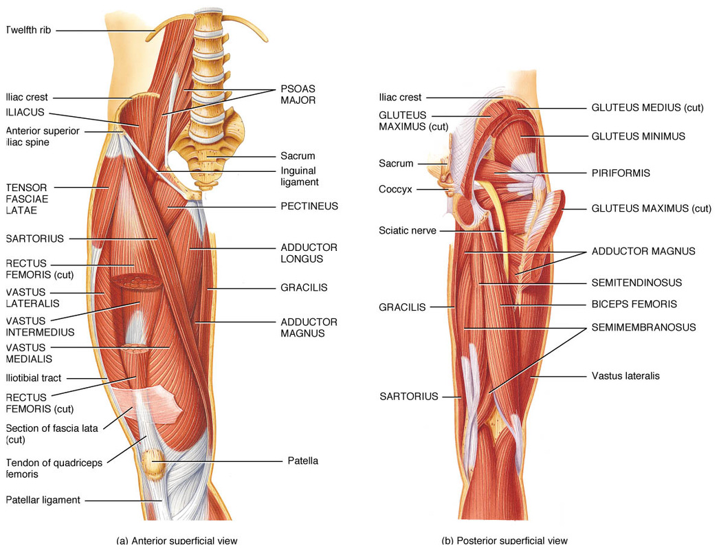
Muscles
The muscles of the thigh and lower back work together to keep the hip stable, aligned and moving. It is the muscles of the hip that allow the 4 basic movements of the hip:
- flexion – bend
- extension – straighten
- abduction – take the leg away from the body
- adduction – bring the leg back toward the body
Muscles of the Hip Region:
- Gluteus Minimus
- Gluteus Medius
- Gluteus Maximus
- Tensor Fascia latae
- piriformis
- Obturator
- Gracilis
- Adductor Muscles
- Abductor Muscles
- Iliopsoas
- Rectus Femoris
- Vastus Lateralis
- Vastus Intermedius
- Vastus Medialis
- Psoas Major
- Pectineus
- Sartorius
- External Rotators
- Hamstring
Nerves (major)
Nerves carry signals from the brainto the muscles to move the hip and carries signals from the muscles back to the brain about pain, pressure and temperature.
- Femoral
- Sciatic
- Obturator
Blood Vessels (Major)
- Femoral Artery
- Internal and External Iliac
- Superior and Inferior Gluteal
Bursae (Major)
Bursae are fluid filled sacs lined with a synovial membrane which produce synovial fluid. The synovial fluid is similar in consistency to raw egg white. Bursae are often found near joints. Their function is to lessen the friction between tendon and bone, ligament and bone, tendons and ligaments and between muscles. There are as many as 20 bursae around the hip. Inflammation or infection of the bursa is called bursitis.(1)
- Greater Trochanteric Bursae
- Iliopsoas Bursae
- Ischial Bursae
References
- 1. http://www.healthpages.org/anatomy-function/hip-structure-function-common-problems/#anatomy-of-the-hip
- http://www.nlm.nih.gov/medlineplus/hipinjuriesanddisorders.html
